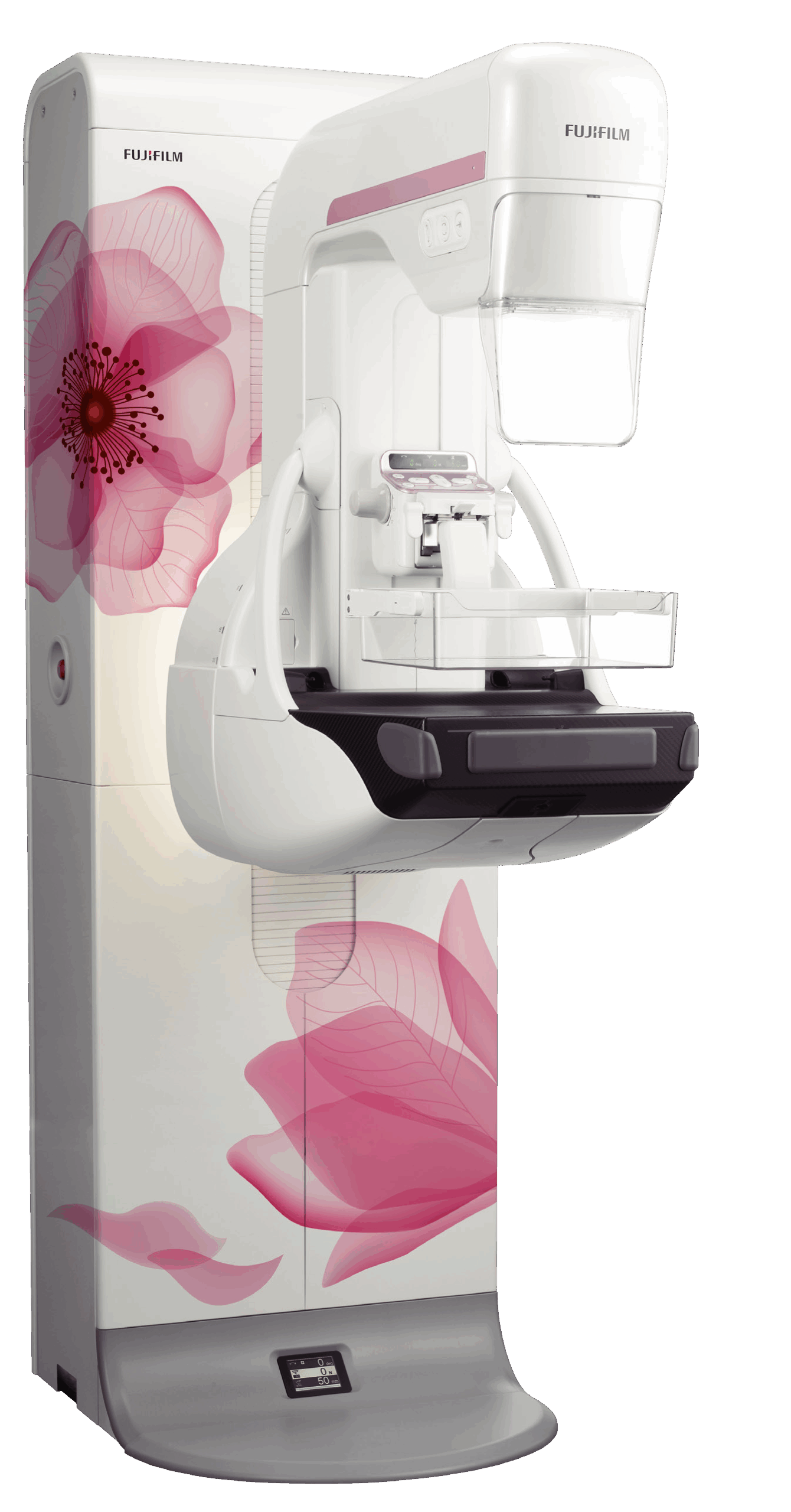
Learn more about the individual tests at DA MedSuites:
- Mammogram
- Ultrasound Breast
Mammograms and ultrasounds breast are both crucial tools in the early detection of breast cancer, but they approach the task from different angles. When used in conjunction, they offer a more comprehensive and accurate picture of breast health, ultimately leading to improved outcomes. Here's why the combination of these two imaging techniques holds significant value:
1. Complementary Technologies:
- Mammograms excel at: Detecting microcalcifications (tiny calcium deposits) and subtle masses. They utilize X-rays to create images of the breast tissue, which can reveal abnormalities that might be missed by palpation (physical examination).
- Ultrasounds excel at: Differentiating solid masses from fluid-filled cysts. They use sound waves to create images, making it easier to assess the characteristics of a breast mass. They are particularly helpful in visualising dense breast tissue, where mammograms may be less effective.
2. Addressing Limitations:
- Mammograms in Dense Breasts: Women with dense breasts (more fibrous and glandular tissue, less fatty tissue) can find their mammograms harder to interpret. Dense tissue can obscure potential tumors, making them harder to spot. Ultrasound can help "see through" this density by providing a clearer view of the underlying structures.
- Ultrasound's Limitations: While excellent at distinguishing solid from fluid-filled masses, ultrasound may struggle to detect microcalcifications, which are often an early sign of cancer. It can also be difficult to visualise deep-seated tumors with ultrasound alone.
3. Improved Detection Rates:
- Studies have shown that the combined use of mammography and ultrasound, particularly in women with dense breasts, leads to a higher rate of early cancer detection compared to mammography alone. This earlier detection can lead to more effective treatment and better survival rates.
4. Personalized Approach to Screening:
- Healthcare providers can tailor screening recommendations based on an individual's risk factors and breast tissue density. For women with dense breasts or other risk factors, a combined approach of mammography and ultrasound is often recommended. This personalized approach optimises the effectiveness of screening efforts.
5. Reducing False Positives:
- While the combined approach can increase detection rates, it also comes with the potential for more false positives (findings that appear suspicious but are ultimately benign). However, in many cases, the additional information gained from ultrasound can help clarify the nature of the abnormality, potentially reducing the need for unnecessary biopsies.
In Conclusion:
Mammograms and ultrasounds are valuable tools in breast cancer screening, and their strengths are amplified when used together. By leveraging their complementary abilities, they offer a more comprehensive and accurate assessment of breast health, leading to improved detection rates, better treatment outcomes, and ultimately, a higher chance of survival. Consulting with a healthcare provider is crucial to determine the most appropriate screening strategy based on individual risk factors and breast characteristics.
Accessing your results:
Your mammography and ultrasound breast images and report will be digitally accessible for review by our medical team. If you were referred, your healthcare provider will receive a comprehensive report promptly, enabling them to discuss the findings and recommendations with you during a follow-up appointment.
Printed copies of the images can be provided upon request (additional charges may apply).

