

By Dr Say Tian Ling is a Medical Doctor at Doctor Anywhere
When navigating proactive breast health, women frequently encounter two primary diagnostic imaging methods: mammograms and breast ultrasounds. Understanding the distinct clinical purposes, mechanisms, and limitations of each modality is essential for making informed healthcare decisions.
In Singapore, breast cancer remains the most common cancer among women. The Ministry of Health (MOH) Singapore outlines specific screening frameworks—such as the national Screen for Life program—recommending regular screening for average-risk women starting from age 50, and individualised clinical consultations for women aged 40 to 49.
What is a Mammogram?
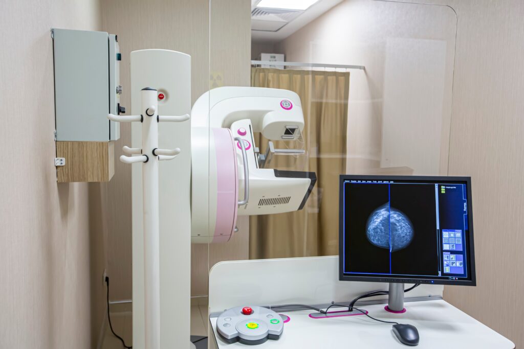
A mammogram is a specialised diagnostic procedure that uses low-dose X-ray imaging to create detailed cross-sectional views of the breast tissue.
- How it Works: During the procedure, the breast tissue is gently compressed between two specialised plates. This flattening minimises tissue thickness, reducing the required radiation dose and optimising image clarity.
- Primary Clinical Utility: Mammograms are the established primary screening standard for early breast cancer detection. They are uniquely capable of identifying microcalcifications—tiny calcium deposits that often serve as the earliest indicator of early-stage ductal malignancies before physical symptoms or lumps can be felt.
- Clinical Considerations: The compression process may cause brief, temporary physical discomfort. Additionally, because both dense breast tissue and potential masses appear white on an X-ray film, mammography can exhibit reduced sensitivity in women with dense breasts.
What is a Breast Ultrasound?
A breast ultrasound is a non-invasive imaging modality that utilises high-frequency sound waves to capture real-time structural data of the soft tissue.
- How it Works: A clear conductive gel is applied to the skin, and a medical professional guides a handheld transducer over the breast. The transducer records the acoustic waves as they bounce off internal structures, generating real-time digital images.
- Primary Clinical Utility: Breast ultrasounds are primarily used as a supplemental diagnostic tool rather than a standalone screening method. They excel at differentiation, allowing clinicians to definitively distinguish between fluid-filled benign cysts and solid tissue masses that require further evaluation.
- Clinical Considerations: Because ultrasounds do not utilize ionising radiation, they are frequently preferred for initial evaluations in pregnant individuals or women under the age of 30 who present with localised symptoms. However, ultrasound technology cannot reliably detect micro-calcifications.
Direct Comparison: Mammogram vs. Breast Ultrasound
The table below outlines the core differences between the two modalities based on clinical applications:
| Diagnostic Feature | Mammogram | Breast Ultrasound |
| Imaging Modality | Low-dose X-ray radiation | High-frequency sound waves (No radiation) |
| Primary Indication | Routine screening for asymptomatic women aged 40+ | Target evaluation of lumps, dense tissue, or young patients. |
| Detection Capabilities | Highly effective for micro-calcifications and deep structural shifts | Differentiates solid masses from fluid-filled cysts |
| Limitations | Reduced sensitivity in highly dense breast tissue | Cannot reliably identify early micro-calcifications |
Which Screening Tool Do You Actually Need?
Clinical guidelines indicate that a breast ultrasound cannot replace a screening mammogram. For the majority of asymptomatic women over 40, the mammogram remains the primary, evidence-based frontline tool for reducing breast cancer mortality.
However, patients often require both modalities simultaneously under the following medical parameters:
- Dense Breast Tissue: If a screening mammogram indicates high tissue density, a supplemental ultrasound provides an added layer of evaluation to view areas potentially obscured on the X-ray.
- Investigating Abnormalities: If a routine mammogram reveals an ambiguous asymmetry or potential mass, a subsequent ultrasound is deployed to evaluate the precise composition of that specific area.
“On a regular mammogram screen, both dense breast tissue and tumors show up as white. This can create a “cloudy” or blurry effect, making it easy for a small lump to hide behind healthy tissue. If your doctor mentions you have dense breasts, adding a simple ultrasound to your regular mammogram gives you the clearest, most complete picture of your health.”
– Dr Say
At DA Orchard MedSuites, our comprehensive women’s health screenings look beyond isolated symptoms to the deeper systems that shape how well, and how long, you live. This kind of evaluation exists because it answers what a standard annual check often doesn’t: how efficiently your body is really functioning beneath the surface, how your metabolic health is adapting, and what’s happening at a cellular level long before it would ever show up as a diagnosis.
Rather than waiting for a number to turn red, it’s a proactive step to help you understand — and manage — your health trajectory early. Book your health screen today at DA Orchard MedSuites here.

